The respiratory and cardiac stage is where auscultation enters the course, and with it the hardest documentation lesson so far: a sound is only a finding when its location, timing and quality are written down, which makes anatomical landmark language the real curriculum of the week. The written work is a documented thorax and heart assessment from your own stethoscope practice. Your section may print this as NR 302 or NR302; it is the same course. Chamberlain publishes no syllabi outside Canvas. The placement here is our teaching judgment from the course's catalog arc; your section's rubric decides what your week actually asks.
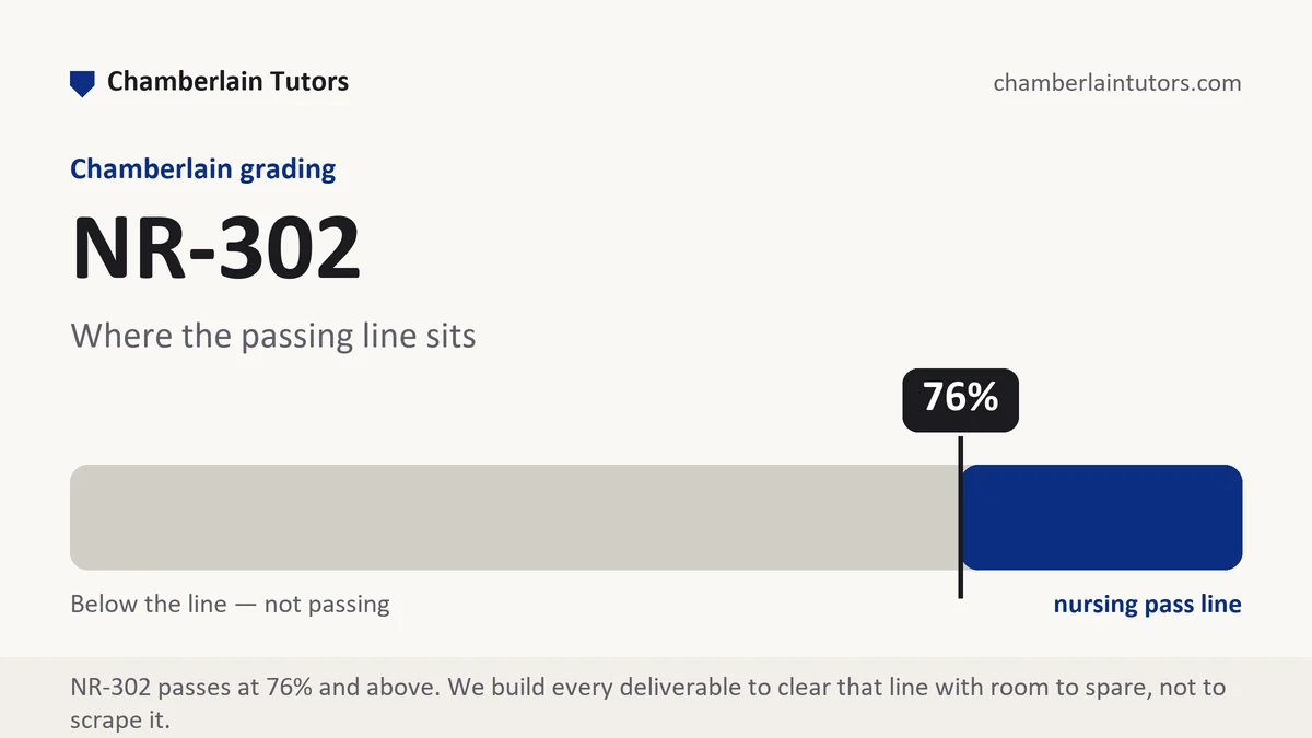
What NR-302 Week 6 asks for
Lungs clear is the two-word entry that tells a chart reviewer almost nothing, and the reviewer's first question exposes why: clear where? A trained respiratory assessment listens at named sites down both sides of the chest, anterior and posterior, comparing side against side, and its documentation carries that geography: breath sounds clear to auscultation bilaterally, anterior and posterior, all lobes. The difference between the two entries is not thoroughness of writing; it is whether the writing proves an exam. The sixth stage of an assessment course brings the stethoscope into the curriculum, and its write-ups are graded on exactly that proof.
The territory divides in two. The respiratory side covers inspection of the chest's shape and breathing effort, the respiratory pattern with its rate and depth, palpation as taught at this level, and auscultation of breath sounds at the systematic sites with their expected qualities. The cardiac side covers the apical pulse with its location described against the chest's landmark lines, rate and rhythm by auscultation, the first and second heart sounds as heard, and the peripheral circulation that completes the picture: pulses graded on the taught scale, capillary refill, and any edema described by location and the assessed scale. Both sides share one grammar: landmark, method, finding, quality.
Deliverables at this stage run to the usual shapes, a documented practice assessment, a lab write-up, a scenario documentation exercise, with one addition worth expecting: the symptom layer grows teeth here, because chest and breathing complaints are the interview material this region owns. A reported breathing symptom run through its full attribute set, and a documented set of pertinent denials, cough, shortness of breath, chest discomfort, palpitations, is scored as seriously as the auscultation entries. As everywhere in this course, hearing something odd in a practice partner is described, never diagnosed, and the write-up's job is a finding the next clinician could relocate with their own stethoscope.
The NR-302 Week 6 method, step by step
Six moves for documenting what the stethoscope and the landmark lines produce.
-
Learn the landmark lines as writing vocabulary first
Midclavicular, midaxillary, the intercostal spaces, the named auscultation areas. Every finding this week is located against that grid, and the write-up that cannot name its location cannot document its finding.
-
Document the respiratory pattern before the sounds
Rate counted for the full taught interval, depth, effort, symmetry of chest movement. The pattern entries frame the auscultation and are the part most often skipped by students eager to write about sounds.
-
Record breath sounds site by site, side against side
The systematic sequence your text teaches, documented as performed: clear bilaterally at all assessed sites, or the deviation at its named site with its quality described in the taught vocabulary.
-
Locate the apical pulse in landmark language
The auscultation point described against the intercostal space and the reference line, with rate counted apically for the full interval and rhythm characterized as regular or with its irregularity described.
-
Write the heart sounds as heard, not as memorized
First and second sounds present and distinct at the assessed areas is a beginner's honest entry. Extra sounds are described by where and when in the cycle they occurred, without naming a suspected cause.
-
Complete the circuit with the periphery
Paired pulses graded on the taught scale per site per side, capillary refill with its timing, edema present or absent by location with the scale if assessed. The cardiac write-up ends at the fingertips and ankles, not at the chest.
A layout and word budget for a thorax and heart write-up
The frame below fits a documented cardiorespiratory assignment of roughly 850 to 1,050 words. It is our own guide rather than anything the university issues, and your week's rubric and assigned form outrank it wherever they disagree.
| Section | What belongs in it | Word target |
|---|---|---|
| Regional symptom layer | Reported breathing and chest symptoms with full attributes, plus the pursued denials, in reporting verbs. | 130 to 160 |
| Chest inspection and pattern | Shape, symmetry, effort, rate with its counting interval, and depth, before any sound appears. | 130 to 160 |
| Breath sounds by site | The systematic sequence with laterality throughout, qualities in the taught vocabulary, deviations located precisely. | 170 to 210 |
| Apical assessment | Location in landmark language, rate for the full interval, rhythm, and the first and second sounds as heard. | 140 to 170 |
| Peripheral circulation | Graded pulses per site per side, refill timing, and edema status by location. | 120 to 150 |
| Method and comparison note | What was assessed by which technique, compared against the cited expected findings, scope held. | 90 to 120 |
Evidence craft for auscultation documentation
Location is the license for every sound entry. A breath sound without a site or an apical rate without its landmark is unverifiable. The intercostal-space-and-line grammar exists so a second listener can place their stethoscope where yours was, which is the entire meaning of documented.
Counting intervals are part of the datum. A rate counted for the full taught interval is a different measurement from a shortcut estimate, and rhythm irregularities make the difference clinically material. Write the interval with the count and the habit becomes visible credit.
Grading scales must be named to mean anything. A pulse graded two means nothing until the scale is identified; the taught scale with its range makes the entry portable. The same holds for edema grading and any quality vocabulary: the textbook's terms, used as defined, cited where comparison is assigned.
Symmetry claims require both sides examined. Bilaterally is a word your exam has to earn: it asserts the sequence visited matching sites on both sides. If a site was inaccessible in the practice session, the honest entry names what was assessed and what was not, exactly as the previous weeks taught.
Five mistakes that cost points in this week's territory
- Lungs clear, unlocated. The two-word entry proves no sequence, no sites and no comparison, and this is the week the habit is priced out of you.
- The radial shortcut for apical work. Documenting an apical rate that was actually taken at the wrist misstates the method, and method is part of the finding.
- Sounds named beyond scope. An unexpected sound described by site, timing and quality is correct; the same sound labeled with a suspected condition is a scope breach.
- Ungraded pulses. Present is not a grade; the taught scale per site per side is the standard, and its absence collapses the peripheral section.
- A soundless symptom layer. Skipping the reported cough, breathlessness and chest-discomfort questions leaves the region's interview half undone, and rubrics check the denials.
Before you submit
- Every sound entry carries a named site and laterality
- Rates state their counting interval; rhythm is characterized
- The apical location is written in landmark language
- All grades name their scales as taught in the course text
- Deviations are described by site, timing and quality, never named diagnostically
- The regional symptom layer includes the pursued denials
Writing up thorax and heart for NR-302?
Send the instructions and the rubric out of Canvas. A premium original draft comes back in 24 to 48 hours with every sound located and every grade on its scale, and revisions run until the grade lands.